Menstuff® has compiled the following information on the use of
color doppler prior to a biopsy for prostate cancer. Google search
for "color doppler" prostate 13,300.
Color Doppler and Tissue Harmonic Ultrasound in the Early Detection and Staging of Prostate Cancer
Over the years, increases in the reported incidence of PC have been disproportionate to the changes in population demographics. The main reason for this rapid rise may be the easy access to PSA and subsequent ultrasound guided biopsies (random biopsy). Ironically though, this presents a dilemma for the patients and the clinicians. Although saving the lives of many men, some men may have so-called “latent” or “insignificant” tumors that may not need any treatment. Precise ultrasound evaluation with proper biopsy will provide us with valuable information to make a decision between watchful waiting and appropriate early intervention.
Gland Volume and Diagnosis
There is much debate about how to use the diagnostic tools that we have – DRE, diagnostic PSA levels, PSA in relation to age, and gland volume – for early detection of PC. In our practice, a serum PSA > 3ng/ml, a PSA increase of 1ng/ml in a year, or abnormal DRE are the indications for TRUS. If a man’s serum PSA is greater than the predicted PSA (gland volume x 0.12), he is in a “high-risk” group for cancer. This group is subjected to careful ultrasound evaluation.
Making the Decision to Biopsy
Sonographic Evaluation
Because clinically relevant (>0.5 cm3) PC is nearly always hypoechoic (black on ultrasound) compared with normal prostate tissue, we only biopsy lesions that are visible by ultrasound.
Depending on tumor architecture, the degree of hypoechogenicity (darkness on ultrasound) ranges from obvious (nodular) to subtle (infiltrative) changes. Thus, it is incumbent on the physician performing the examination to be familiar with the zonal anatomy and morphologic presentation of prostate cancer.
Cancers in the outer gland (peripheral zone and central zone) and inner gland (transition zone) have different sonographic appearances and biologic behavior, and our threshold that defines whether to biopsy varies depending on lesion size, location, and amount of excess PSA.
Photo here with chart
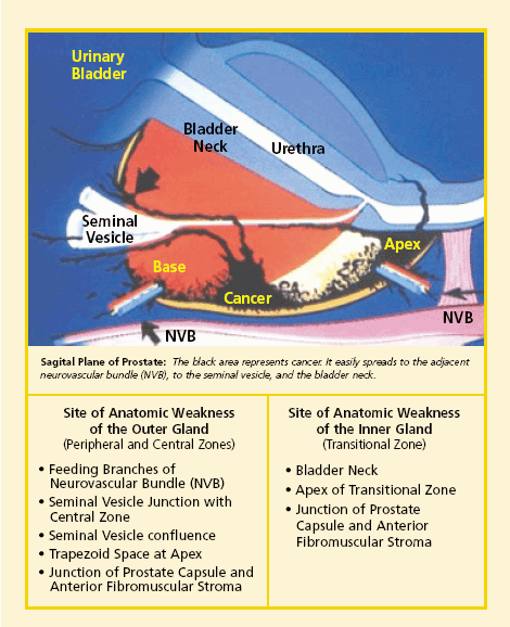
Outer Gland Cancers
Outer gland cancers have a greater propensity than inner gland cancers for extracapsular spread because they can escape easily through the area of anatomic weakness (entry of neurovascular bundle branches, seminal vesicles, and apex). Fortunately, these tumors are easy to visualize because the background tissue is more homogeneous than that of the inner gland. Most outer gland cancers originate laterally at the entrance of the neurovascular bundles. To visualize and sample this area, we have found it best to perform the scanning and biopsy in the transverse plane. When targeting outer gland lesions, we first biopsy the lesion and then sample the accompanying neurovascular branches tangentially along a plane just external to the prostatic capsule. A finding of a tumor intermixed with fat definitively diagnoses histologic stage T3 cancer.
When outer gland tumors extend to the midline, we perform a biopsy of the confluence of the seminal vesicle and trapezoid space of the apex. The base and apex of the gland in this area are always biopsied to aid in the evaluation of the internal spread of cancer.
Hypoechoic lesions of the outer gland should be pursued vigorously because they can escape when they are relatively small. For this reason, we generally perform a biopsy of the lesions we see on the ultrasound when excess PSA suggests that a 1cm3 lesion may be present (excess PSA greater 2 ng/ml).
If we do not find lesions in the outer gland by ultrasound, we generally do not perform random biopsies. At this point, we shift our attention to the inner gland (transition zone).
Inner Gland Cancers
TRUS can detect cancers in the inner gland, though its sensitivity is less than that for the outer gland. If the excess PSA is 4 to 6 ng/ml and no lesion is found in the outer gland, one must carefully scan the inner gland for a homogeneous, poorly defined hypoechoic lesion. We focus on the sites of anatomic weakness of the inner gland, the anterior apex and the bladder neck. Color-flow Doppler and (lately) Tissue Harmonic aid in the diagnosis of these more difficult-to-see inner gland cancers because most tumors larger than 1 cm3 have neovascularity (abundant vessel inside of tumor) that is easily identifiable with these new technologies. Given the confusing heterogeneous nature of the transition zone, color-flow may be the only clue for the presence of cancer in a subtle hypoechoic lesion.
For the inner gland, we take a watchful waiting approach when (1) gland volume is greater than 50 cm3, (2) no suspicious lesion of the anterior apex or bladder neck area is seen, (3) excess PSA is less than 4 to 6 ng/ml, and (4) there is no outer gland lesion. In general, inner gland cancers have less aggressive prognostic factors (Gleason score and DNA ploidy) than outer gland cancers and tend to be confined until they attain very large volumes. Therefore, we feel that these cancers do not need to be pursued as aggressively as outer gland cancers. To ensure that we have not overlooked a significant tumor, we repeat serum PSA testing at 4- to 6-month intervals. Should an upward trend continue, we re-ultrasound.
Staging Biopsy Technique
The biopsy samples should include one sample from the middle of the lesion, and all routes of possible tumor escape based on known sites of anatomic weakness. The positive neurovascular bundle biopsy has to include fat cells in contact with tumor cells or the invaded nerve sheath; a seminal vesicle biopsy should include pigmented epithelium (specific cell layer of seminal vesicle). Because the prostate gland does not contain fat, the presence of this tissue in the specimen confirms an extraprostatic invasion. We stain the rectal end of the tissue core with blue ink before sending it to the laboratory. This will allow us to determine the exact location of the tumor – an inked end signifies an outer gland (peripheral zone) tumor and a non-inked end tumor indicates an inner gland tumor (transition zone).
USING TRUS in PC
Staging and Diagnosis
Information (Risk Factors) Needed from TRUS and Staging Biopsy
What is the ploidy of the tumor?
This information will provide the exact local staging of the cancer and will thereby help the physician and patient choose appropriate a further staging work-up and decide on eventual treatment options.
State-of-the-Art Ultrasound Equipment
It is important to use a high-end up-to-date ultrasound unit for an early detection and accurate staging biopsy. Power Color Doppler ultrasound demonstrates all the blood flow patterns inside the prostate. Usually, cancer tissue shows a higher blood flow (tumor neovascularity) than that of normal tissue. This capability will improve detection and actual tumor size measurement.
The newly developed Tissue Harmonic technology improves spatial resolution to permit visualization of smaller objects and improves contrast resolution to discern very subtle differences in grayscale. This is different from conventional ultrasound imaging, which sends out a burst of sound and listens for that burst to echo off structures in the body, (an echo that is usually weak and distorted). The time it takes for the echo to return is proportional to the distance the sound wave traveled. In Tissue Harmonic technology, instead of listening for the same sound burst to return in the echo, the ultrasound equipment listens only for a sound burst at twice the transmitted frequency. Good ultrasound evaluation with staging (strategic) biopsy may eliminate an unnecessary endorectal MRI study (that is still an imaging study without tissue confirmation). Moreover, it will eliminate the “guesstimation” from random biopsies. Currently, we use the Hitachi EUB-6500 Ultrasound model. Soon, there will be further developments in TRUS that will include contrast (IV form of micro-bubbles), enhanced Color Doppler, and three-dimensional imaging capability.
The Role of TRUS-Guided (Not Random) Biopsies in Determining the Internal and External Spread of PC
Dr. Fred Lee and I published our data comparing sextant (random) biopsy proven PC data with our staging biopsy data on 110 men. All men came to us for a second opinion with known cancer. We performed TRUS with repeat staging biopsies on all of those men. (Seminars in Urologic Oncology, Vol 16, 1998, p 129-136.)
The results were as follows:
Conclusion
Current methods for determining confined PC for the individual patient are only guesstimations. The pathological outcomes for clinically confined PC have only a 50% probability of being correct. Today’s patients seek answers through patient advocacy groups, Internet surfing, and scientific literature. When one of our patients consults with the “specialists”, he quickly surmises their uncertainty. In our hands, the use of state-of-the-art TRUS with Color Doppler and Tissue Harmonic has helped us and others resolve the uncertainty of whether a cancer is or is not confined and what other risk factors they may have. Then, and only then, do we more reliably predict a prognosis and guide our patients to those treatments that are most appropriate for them.
References
McNeal J: Cancer volume and site of origin of adenocarcinoma in the prostate: Relationship to local and distant spread. Hum Pathol 23:258-266, 1992
Lee F: Prostate cancer: Transrectal ultrasound and pathology comparison. Cancer 67:1132-1142, 1991
Epstein J. Corellation of prostate cancer nuclear deoxyribonucleic acid, size, shape and gleason grade with pathological stage at radical prostatectomy. J Urol: 148:87-91, 1992
Bostwick D. Optimized microvessel density analysis improves prediction of cancer stage from prostate needle biopsies. Urology 48: 47-57, 1996
Lee F. Bahn D. The role of TRUS-guided biopsies for determination of internal and external spread of prostate cancer. Seminars in Uro Oncology 16: 129-136, 1998
Source: Prostate Cancer Research Institute
(PCRI), www.prostate-cancer.org/education/staging/Bahn_ColorDopplerUltrasound.html
![]()
Endorectal Color Doppler Sonography and Endorectal MR Imaging
Features of Nonpalpable Prostate Cancer
F. Cornud1, K. Hamida1, T. Flam2, O. Hélénon1, Y. Chrétien2,3, N. Thiounn2, J. M. Correas1, J. M. Casanova4 and J. F. Moreau1
1 Service de Radiologie, Hôpital Necker, 149 rue de Sèvres, 75015 Paris, France.
2 Service d'Urologie, Hôpital Cochin, 24 Rue du Faubourg saint Jacques, 75014 Paris, France.
3 Service d'Urologie, Hôpital Necker, 75015 Paris, France.
4 Service d'Uro-Gynécologie, Hôpital Notre Dame de Bon Secours, 14 rue des volontaires, 75014 Paris, France.
OBJECTIVE. The purpose of this study was to describe endorectal sonography and color Doppler sonography features of nonpalpable prostate cancer and to assess the value of endorectal MR imaging for the preoperative local staging of these tumors.
MATERIALS AND METHODS. Ninety-four patients with nonsuspicious findings on digital rectal examination and a mean prostate-specific antigen level of 16.3 ± 10 ng/mL (median, 13 ng/mL) underwent endorectal sonography, color Doppler sonography, sextant endorectal sonographically guided biopsy, and endorectal MR imaging before radical prostatectomy.
RESULTS. Tumors were visible in 48 cases and not visible in 46. The mean Gleason biopsy score, the frequency of tumors involving three sextants or more of the prostate gland at biopsies, and the frequency of stage pT3 tumors were significantly higher in patients with visible tumors (5.9 ± 0.9, 42%, and 37.5%) than in those with invisible tumors (5.4 ± 1.1, 17%, and 17%). The 42 hypervascular tumors were hypoechoic in every case and had a higher rate of Gleason tumor grades 4 and 5 at biopsy than did the 52 hypovascular tumors (33% versus 11.5%). Six hypovascular tumors (6/52, 11.5%, two visible) had an insignificant tumor volume. Established extraprostatic tumor spread was detected on MR imaging in six of 18 cases (sensitivity, 33%; specificity, 100%0, all of which had the following four features: hypervascularity, prostate-specific antigen level greater than 20 ng/mL, three or more sextants of the gland having positive findings at biopsy, and seminal vesicle invasion.
CONCLUSION. Endorectal sonography and color Doppler sonography are useful to differentiate low-risk invisible and hypovascular tumors from high-risk visible and hypervascular tumors. However, MR imaging has a poor sensitivity for the detection of extraprostatic spread and is accurate only in a minority of highly selected high-risk hypervascular tumors.
American Journal of Roentgenology,
http://www.ajronline.org/cgi/content/abstract/175/4/1161
![]()
Prostatic cancer: role of color Doppler imaging in transrectal
sonography
Department of Medical Imaging, St. Frances Xavier Cabrini Hospital, Malvern, Victoria, Australia.
OBJECTIVE: The aim of this study was to assess the roles of
transrectal color Doppler and gray-scale sonography in revealing
prostatic cancer, using biopsy as the reference standard. SUBJECTS
AND METHODS: Two hundred fifty-six patients referred for urologic
studies underwent transrectal sonography using gray-scale and color
Doppler scanning. All abnormal areas shown on gray-scale or color
Doppler sonography or both were targeted and biopsies were performed.
The patients also underwent random sextant biopsies. All biopsies
were individually correlated with histopathologic findings and all
results were analyzed. RESULTS: Cancer was found on biopsy in 100
patients (39%), and equivocal sonographic results or prostatic
intraepithelial neoplasia was found in 22 other patients (9%). In 16
of the patients in whom cancer was detected, the tumors were
correctly revealed only with color Doppler sonography. These 16
patients had a mean Gleason score of 6.4 (range, 5-8). Biopsy
findings in these 16 patients showed eight patients with extensive
lesions, three with moderate lesions, and five with minimal lesions.
However, in nine other patients with cancer (9% of cancers detected),
both gray-scale and color Doppler sonography failed to reveal lesions
that were found on sextant biopsy. An analysis showed that, although
highly sensitive, color Doppler sonography was somewhat less specific
than gray-scale sonography. CONCLUSION: Color Doppler sonography
should become a routine part of transrectal sonography of the
prostate gland to improve detection and targeting of lesions. The
practice of performing random sextant biopsies should also
continue.
Source: American Journal of
Roentgenology,
www.ajronline.org/cgi/content/abstract/171/1/205
![]()
Technology Improves Prostate Cancer Detection
The information offered in the study is "very important," says Haakon Ragde, MD, clinical assistant professor of urology at the University of Washington Medical School in Seattle, clinical professor at the University of Virginia in Charlottesville, and co-founder of the Pacific Northwest Cancer Foundation.
Few Doctors Are Skilled in the Technology or Have the Equipment
But, Ragde cautions, the utility of the finding is limited to the small percentage of urologists and oncologists who are highly skilled in ultrasound.
The advanced method relies on several ingredients: a newer ultrasound image enhancer, called a microbubble ultrasound contrast agent; a color Doppler ultrasound instrument; and, a practitioner who is experienced in interpreting what appears on the screen.
The new contrast agents produce better signals and bubbles tiny enough to enter small blood vessels, explains the study’s principal investigator Ferdinand Frauscher, MD. Frauscher is a research fellow in the Ultrasound Research and Education Institute of Thomas Jefferson University in Philadelphia and head of the uroradiology division at the University of Innsbruck in Austria. The study of 84 patients with an average age 57 was begun in Innsbruck and completed in Philadelphia.
New Method Tested Against
Old
Each patient was biopsied by two investigators, using different methods. The first investigator took five or fewer samples, guided by the microbubbles and the color Doppler imaging. Tumors generally grow faster than healthy tissue, the authors write, and have increased blood flow. The signals from these areas of increased blood flow determined the placement of the biopsies.
The second investigator took 10 biopsies, systematically taking samples from the different regions of the prostate. Standard or gray-scale ultrasound guided the investigator around the prostate but did not pick up tumors. Six to 12 biopsies are commonly taken in the standard method.
The result: With five or fewer biopsies, the contrast-enhanced color Doppler detected prostate cancer in 23 of 84 patients (27%). They included seven patients (8%) whose random biopsies from the standard method were negative.
With 10 biopsies, the standard method detected cancer in 17 of these same patients (20%), including one patient whose cancer was detected only by the standard method, not the color Doppler. Such cancers tend to have less blood flow than those with higher scores, explaining why it was not detected by the color Doppler method. And, the researchers note, such cancers also tend to grow very slowly and are rarely fatal.
"We are happy because we have improved the detection rate while decreasing the number of biopsies, and that’s very important," says Frauscher.
Needed: FDA Approval and More Experienced Clinicians
A major stumbling block to wide use of this technique in the US, outside of research, is lack of US Food and Drug Administration (FDA) approval for contrast agents now used with great effect in Europe, Japan, and Australia, says Flemming Forsberg, PhD, associate professor of radiology and head of research at the Jefferson Ultrasound Institute at Thomas Jefferson University in Philadelphia.
"At the end of the day," Forsberg says, "the honest, ugly truth is that with [standard] transrectal ultrasound, we only find 55 to 60 percent of cancers, which is marginally better than flipping a coin and saying, ‘You’ve got it, you haven’t got it.’" That is why several random biopsies are currently done in men suspected of having prostate cancer.
The FDA has approved one newer contrast agent, Optison, for use in diagnosis of heart disease; clinicians could use it as an "off-label" product for prostate color ultrasound, says Frauscher, who thinks it prudent to wait for the official FDA sanction.
Radiologists are eager for approval because the new agents have the potential for making a major difference in diagnosing prostate cancer, Forsberg says, as doctors gain training and skill in their use.
"I’m not thinking that color and bubbles will necessarily show you everything there is," Forsberg says. "What I think this does is allow you to do your regular six or 12 biopsies, and then add some color-directed biopsies to areas of suspicion you detect. At the same time, I’m not thinking this will give you less needles. But I think it will get you the right needles."
Early Detection Needed for Successful Treatment
The delay in FDA approval has concerned ultrasound and radiology professionals so much that two professional associations have formed a joint task force, with the cooperation of competing pharmaceutical companies, to review confidential communications between the FDA and the companies, trying to get a picture of the FDA’s issues, Forsberg says. Soon, the task force will request a meeting with the FDA.
Ragde, the Seattle urologist, firmly believes patients with rising PSA levels and negative biopsies should seek well-experienced practitioners of the color Doppler method — FDA approval or not. Ragde led the first team in the US that treated prostate cancer with ultrasound-guided radiation seed implants, and he also has studied the effectiveness microbubble contrast agents.
"It’s very important to find early cancers by whatever method
you find them," Ragde says, "because earlier cancers can be
cured."
Source: American Cancer Society, www.cancer.org/docroot/NWS/content/NWS_1_1x_Technology_Improves_Prostate_Cancer_Detection.asp?sitearea=NWS&viewmode=print&
![]()
Enhanced Ultrasound Improves Detection of Prostate Cancer
Prostate cancer is a common cancer among men in the United States. Prostate cancer is the second leading cause of cancer death in men in the United States. The prostate is a walnut-size gland that is located between the bladder and rectum and forms a component of semen. Prostate specific antigen (PSA) levels (a protein produced by the prostate that is elevated when cancer is present), a digital rectal exam (DRE) and transrectal ultrasound are common tests used to detect prostate cancer. If any suspicious mass is found through these tests, a patient must then undergo biopsies (the removal of a sample of tissue) to definitively determine whether cancer exists. However, it is imperative that a physician takes a biopsy from the area in the prostate where the cancer exists to provide accurate diagnostic information. Physicians often use endorectal ultrasound to help determine where in the prostate to take a biopsy. Researchers are attempting to improve upon the accuracy of ultra sound in the guidance of placement of biopsies, including the introduction of contrast, which help physicians to discern between healthy looking tissue and possible sites of cancer.
A new development in ultrasound involves the use of color Doppler imaging with microbubble contrast so that physicians are better able to determine the presence and exact location of a mass within the prostate. Doppler imaging can sense differences in velocity (i.e. blood flow versus solid tissue) and transmits these differences through different color pixels to create a picture on a screen. Microbubbles are tiny bubbles of gas that can permeate through small blood vessels without creating any harm. The microbubbles further enhance imaging by increasing the intensity of backscatter signal. Since blood vessels and blood flow are more prevalent in cancerous tissues than regular tissues, microbubbles tend to concentrate in the cancer, which is revealed on the created picture. This allows physicians to more accurately locate where biopsies should be taken.
Researcher from France recently conducted a clinical study to determine the effectiveness of contrast-enhanced color Doppler ultrasound using microbubbles in determining biopsy sites in men suspected of having prostate cancer. This trial included 85 men who underwent conventional Doppler and microbubble-enhanced color Doppler during the biopsy procedure. The results between the two were directly compared based on biopsy results. Contrast-enhanced color Doppler had a 93% detection rate of prostate cancer, compared with only 54% for un-enhanced color Doppler. Biopsies from areas of the prostate that did not contain cancer occurred in 21% of biopsies under Doppler that was not enhanced, compared with only 11% of biopsies under contrast-enhanced Doppler.
The researchers concluded that microbubble-enhanced color Doppler used for endorectal ultrasound improves the detection of prostate cancer and reduces unnecessary biopsies, compared to color Doppler that is not enhanced. They also state that this procedure is simple and not time consuming. Patients suspected of having prostate cancer may wish to speak with their physician about the risks and benefits of microbubble-enhanced color Doppler in endorectal ultrasound for biopsy placement or the participation in a clinical trial evaluating other novel screening approaches.
Reference: Roy C, Buy X, Lang H, et al. Contrast enhances color
Doppler endorectal sonography of the prostate: efficiency for
detecting peripheral zone tumors and role for biopsy procedure. The
Journal of Urology. 2003;170:69-72
Source: www.youngagain.com/ultrasound.html
![]()
3-D Color Doppler Ultrasound
For men, an ultrasound of the prostate gland may be ordered if a blood test result shows an elevated level of PSA (Prostate Specific Antigen) or if a physician feels a nodule during a patient’s routine physical exam. Ultrasound may also be used to detect other conditions, including inflammation of the prostate or possible reasons for infertility.
Because ultrasound provides real-time images, it can also be used to guide procedures such as needle biopsies - a needle is used to sample cells from an abnormal area for laboratory testing.
The Texas Prostate Institute utilizes rare and highly sophisticated ultrasound equipment that generates anatomic images in three dimensions, providing highly detailed images for use in diagnosis and treatment related to the prostate.
Texas Cancer Clinic, 9102 Floyd Curl, San Antonio, Texas 78240,
888-748-0210, Texas Cancer Clinic, http://www.texascancerclinic.com/tools/print/default.aspx
Glossary
of Prostate Cancer Related Terms ![]() www.prostate-cancer.org/resource/gloss_c.html
www.prostate-cancer.org/resource/gloss_c.html
![]()
Test Overview
A Doppler ultrasound test uses reflected sound waves to evaluate blood as it flows through a blood vessel. It helps doctors evaluate blood flow through the major arteries and veins of the arms, legs, and neck. It can show blocked or reduced blood flow in the arteries of the neck that could cause a stroke. It can also reveal blood clots in leg veins that could break loose and block blood flow to the lungs (pulmonary embolism).
During duplex Doppler ultrasound, a handheld instrument (transducer) is passed lightly over the skin above a blood vessel. The transducer sends and receives sound waves that are amplified through a microphone. The sound waves bounce off solid objects, including blood cells. The movement of blood cells causes a change in pitch of the reflected sound waves (called the Doppler effect). If there is no blood flow, the pitch does not change. Information from the reflected sound waves can be processed by a computer to provide graphs or pictures that represent the flow of blood through the blood vessels. These graphs or pictures can be saved for future review or evaluation.
The four types of Doppler ultrasound are:
Why It Is Done
Doppler ultrasound is done to:
How To Prepare
Nicotine causes blood vessels to constrict; therefore, you may be asked to avoid products that contain nicotine (cigarettes, chewing tobacco) for 30 minutes to 2 hours before the test.
How It Is Done
You will need to remove any jewelry that might interfere with the Doppler ultrasound scan. You will need to take off all or most of your clothes, depending on which area is being examined (you may be allowed to keep on your underwear if it does not interfere with the test). You will be given a cloth or paper covering to use during the test.
Gel is applied to the skin to promote the passage of the sound waves. The transducer is placed in the gel and moved along the skin. You need to lie very still during the procedure. You may hear sounds that represent the flow of blood through the blood vessels.
Arteries in the arms and legs
This test is often performed on both arms or both legs. Even if the suspected blood flow problem is in only one limb, both may be tested for comparison. If your arms are being tested, they will be tested first while you are lying down and then again while you are sitting.
Depending on which blood vessels are being tested, a blood pressure cuff may be wrapped around one or both limbs so the blood pressure can be taken at several different places. When testing the legs, a blood pressure cuff may be wrapped first around the calf and then around the thigh. The test may be done at several locations on your leg. When testing the arms, the pressure cuff may be wrapped first around the forearm and then around the upper arm.
Veins in the arms and legs
For this test, you will be asked to lie down and breathe normally. You must lie very still. Any changes in blood flow that occur as a response to your breathing patterns are noted.
The test may be repeated while the examiner presses on the veins close to the surface of your skin to help detect a clot in the vein (called a compression maneuver). The examiner may do this with your legs or arms in different positions to ensure that the blood supply is not blocked in these positions. The examiner may also squeeze your calf or forearm to help blood move more quickly through the veins (called an augmentation maneuver). This is done to evaluate blood flow toward your heart.
While your legs are being tested, you may also be asked to try to breathe out strongly with your nose pinched and your mouth closed (called Valsalva's maneuver). This maneuver usually causes a sudden change in blood flow.
Arteries in the neck
You will be asked to lie down with a pillow underneath your head for support. The test is performed on both sides of your neck, and then the results are compared to standard values to determine the amount of blockage or narrowing of the arteries.
Transcranial ultrasound
For a transcranial ultrasound, the transducer is passed lightly over the skin at the base or side of your skull.
How It Feels
There is normally no discomfort involved with having a Doppler ultrasound test. The gel may feel cold when it is applied to your skin unless it is first warmed to body temperature. If your blood pressure is taken during the test, you will feel pressure when the blood pressure cuffs are inflated.
Risks
There are no known risks associated with a Doppler ultrasound test.
Results
Doppler ultrasound Normal:
For continuous wave or duplex Doppler, the signals obtained are the same for both sides of the neck or for both limbs. No blockage, aneurysm, or narrowing of a blood vessel (stenosis) is detected. The rate of blood flow is similar to standard values. These results indicate open blood vessels with normal blood flow.
On a duplex Doppler ultrasound graph:
For color Doppler, the image indicating blood flow is normal for both sides of the neck or for both limbs.
Abnormal:
For continuous wave Doppler or duplex Doppler, differences in blood flow between the right and left sides of the body may be heard. At the exact location where an artery is blocked or narrowed, the sound may be high-pitched or turbulent. Blockage (such as from a blood clot), aneurysm, or narrowing of a blood vessel may be detected. The speed of blood flow may be compared to standard values to determine the amount of blockage or narrowing of the blood vessel.
A duplex Doppler ultrasound graph may show irregular flow that indicates a blocked or narrowed blood vessel.
A color Doppler image may show a blocked or narrowed blood vessel or an aneurysm.
In the veins, a blood clot may be indicated if blood flow does not change in response to breathing or does not increase in response to an augmentation maneuver or Valsalva's maneuver. Incomplete blockage of a vein by a blood clot may be seen on color Doppler or during a compression maneuver.
What Affects the Test
Bones above the area being studied or gas in the intestines can interfere with Doppler ultrasound results, because the sound waves do not travel well through bone or gas.
The results of a Doppler ultrasound may not be accurate if you cannot remain still during the test.
Extreme obesity may interfere with the quality of the ultrasound picture.
Irregular heart rhythms (arrhythmias) or heart disease may cause changes in blood flow patterns, even though the blood vessels are not abnormal.
A cold arm or leg that is scanned may give inaccurate results, because blood flow through that limb may be slowed.
Having an open wound in the area that needs to be viewed may interfere with having an ultrasound test.
What To Think About
Producing accurate test results with Doppler ultrasound requires a skilled examiner. The scans should be interpreted immediately in case repeat tests are needed.
Because Doppler ultrasound requires a person to hold very still, some children may need to be sedated so their movements do not interfere with the results.
Angiography and venography are X-ray tests that require the injection of contrast material. In many cases, Doppler ultrasound may be done instead of angiography or venography, since it is faster, less expensive, and noninvasive.
Angiography or venography may be done if results from a Doppler ultrasound are inconclusive. Angiography is usually more accurate than Doppler ultrasound and is considered the most definitive test for evaluating blood flow through an artery. Magnetic resonance angiography (MRA) and CT angiography may be done instead conventional angiography, because these tests are less invasive and easier to perform than conventional angiography. In some cases, venography may be needed to confirm a suspected vein problem. For more information, see the medical tests Angiography and Venography.
Credits
Author Renée Spengler, RN, BSN Source:
my.webmd.com/hw/health_guide_atoz/hw4477.asp?printing=true
![]()
The four types of Doppler ultrasound are:
Resources: Prostae Institute of America, 168 N Brent St,
Ste 402, Ventura, CA 93003 805.585.3082 or 888.234.0004
dvaughan@cmhhosptial.org
Color Doppler Imaging of the Prostate: Important Adjunct to Endorectal Ultrasound of the Prostate in the Diagnosis of Prostate Cancer.
Ultrasound Quarterly. 17(3):185-189, September 2001. Cheng, Sam M.D. *; Rifkin, Matthew D. M.D. +
Abstract:
Summary: The purpose of this article is to evaluate color Doppler imaging (CDI) as an adjunctive tool to gray-scale ultrasound (US) in the diagnosis of prostate cancer and to correlate CDI-positive lesions to cancer grade. We retrospectively analyzed 619 consecutive patients who underwent prostate US, CDI, and biopsy because of abnormal digital rectal examination results or prostate-specific antigen levels. All had directed (into a specific lesion) biopsies or directed biopsies along with systematic four-quadrant or sextant biopsies, or systematic biopsy alone. Color Doppler imaging was compared with gray-scale findings and histologic results. There were 222 (35.9%) biopsy-proven cancers (n = 197) or prostatic intraepithelial neoplasia (n = 25). Of these, 106 (47.7%) had color-flow abnormalities. Of these 106 patients, 26 (24.5%), or 11.7% of all cancer patients, had relatively normal gray-scale US findings but had focal CDI abnormalities as the method of identification. Overall, 76.9% of these were moderate to high Gleason grades and were considered clinically significant lesions. Color Doppler imaging can identify a large number (11.7%) of clinically significant prostate cancers that are poorly seen by gray-scale US. Positive lesions on CDI are of clinical importance because 76.9% are histologically, moderately, or poorly differentiated. We recommend that CDI be used in all diagnostic and biopsy-guided US examinations of the prostate.
(C) 2001 Lippincott Williams & Wilkins, Inc.
www.ultrasound-quarterly.com/pt/re/ultrasoundq/abstract.00013644-200109000-00008.htm;jsessionid=CqP80lhQKiMXoXipmO2RyLBLg1kkKK5H2AkEyOI2tzh7Nutx6kh7!-2128958162!-949856032!9001!-1
![]()
Prostate: techniques, results, and potential applications of color Doppler US scanning.
Rifkin MD, Sudakoff GS, Alexander AA. Department of Radiology, Albany Medical College, NY 12208.
Color Doppler ultrasound (US) scanning and conventional endorectal
gray-scale US of the prostate were performed in 619 patients.
Pathologic correlation was available in all cases after US-guided
transrectal biopsy. There were 132 cancers in 121 men, 13 foci of
atypia in 10 men, 33 foci of inflammation in 31 men, and 469 benign
lesions in 457 men. Two hundred seventy patients with abnormal areas
of flow identified at color Doppler scanning also underwent spectral
waveform analysis of the area of potential concern. No statistical
difference in the mean resistive indexes was identified in any
patient (P = .25; Scheffe F test, analysis of variance). All
malignant lesions had abnormalities demonstrated at gray-scale US
and/or focal or diffuse abnormal flow demonstrated at color Doppler
scanning. Of the 132 cancers, 123 (93%) had corresponding gray-scale
abnormalities and 114 (86%) demonstrated abnormal flow at color
Doppler imaging. Nine of the 132 cancers (7%) had no obviously
identifiable abnormality at gray-scale scanning but had distinctly
abnormal flow at color Doppler scanning. Abnormal findings at color
scanning without abnormal findings at gray-scale scanning occurred in
eight of the 33 cases of inflammatory foci (24%) and in 24 of the 469
(5%) benign lesions
Source: National Library of Medicine,
www.ncbi.nlm.nih.gov/entrez/query.fcgi?cmd=Retrieve&db=PubMed&dopt=Abstract&list_uids=93134064
![]()
Contrast Enhances Color Doppler Endorectal Sonography of
Prostate: Efficiency for Detecting Peripheral Zone Tumors and Role
for Biopsy Procedure
Abstract:
Purpose: We evaluated the accuracy of contrast enhanced color Doppler endorectal ultrasound to guide biopsy for the detection of prostate cancer.
Materials and Methods: A total of 85 patients were evaluated with gray scale and color Doppler before and during intravenous injection of ultrasound contrast agent made of galactose based air microbubbles. Our biopsy protocol was performed during contrast injection. An additional 18 directed cores were obtained based on contrast enhanced imaging. Diagnostic efficiency with and without contrast medium injection for detecting prostate cancer were compared based on biopsy results.
Results: Cancer was identified in a total of 58 biopsy sites in 54 patients. Gray scale imaging revealed 96 abnormal hypoechoic nodules or irregular zones inside the outer gland, of which 48 were malignant on pathological evaluation. Contrast enhanced color Doppler had higher sensitivity (93%) than unenhanced color Doppler (54%), while specificity increased only 79% to 87% for enhanced imaging. Nine of 10 isoechoic suspicious zones were depicted with enhancement, while unenhanced Doppler detected 7 of them. There was no significant difference between the intensity of enhancement and tumor Gleason scores.
Conclusions: Contrast enhanced color Doppler endorectal sonography increases the detection of prostate cancer. Improvement in sensitivity was high, while the difference in specificity was not as pertinent. It is accurate when using a common and routine application ultrasound unit. This technique is easy to perform and not time-consuming. Obtaining additional biopsy cores of suspicious enhancing foci significantly improves the detection rate of cancer.
Source The Journal of Urology, Journal of Urology.
170(1):69-72, July 2003. www.jurology.com/pt/re/juro/abstract.00005392-200307000-00015.htm;jsessionid=CqTlOiv69Z53Yp0Je2glC3Jzg3NefJfhIWEdL1DRTJhFNm72f1J4!-2128958162!-949856032!9001!-1
![]()
![]()
|
|
|